Qlaris Bio came to XVIVO in need of an animation that could visualize the dynamic processes regulating the pressure inside the eye, as well as the mechanisms of lowering this pressure to treat diseases. Certain diseases of the eye, such as glaucoma, are associated with increased pressure inside the eye, which can lead to irreversible blindness. Worldwide, an estimated 76 million adults have some form of glaucoma, and remains a leading cause of blindness.
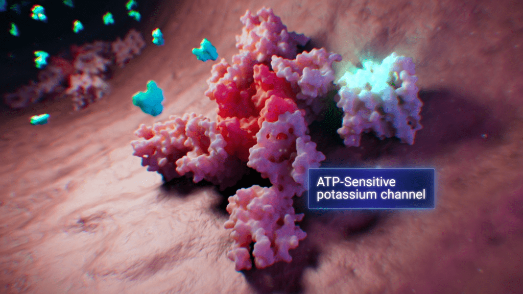
Qlaris Bio is investigating a novel type of medicine to reduce the pressure inside the eye, with the hopes of providing a new and targeted option to people suffering from glaucoma. To help viewers understand how their investigational drug QLS-101 works, the first step was to make the complex anatomy of the eye more understandable and explain the variety of factors that regulate the pressure inside the eye under normal circumstances.
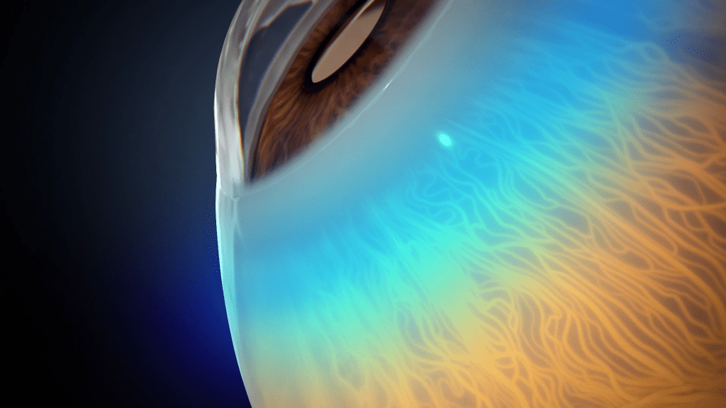
The animation presents fine visual detail of the eye anatomy, including the sinuous path of the episcleral veins and the cross-sectional view of the retina that so closely resembles human anatomy, you would think you were examining someone’s actual eye.
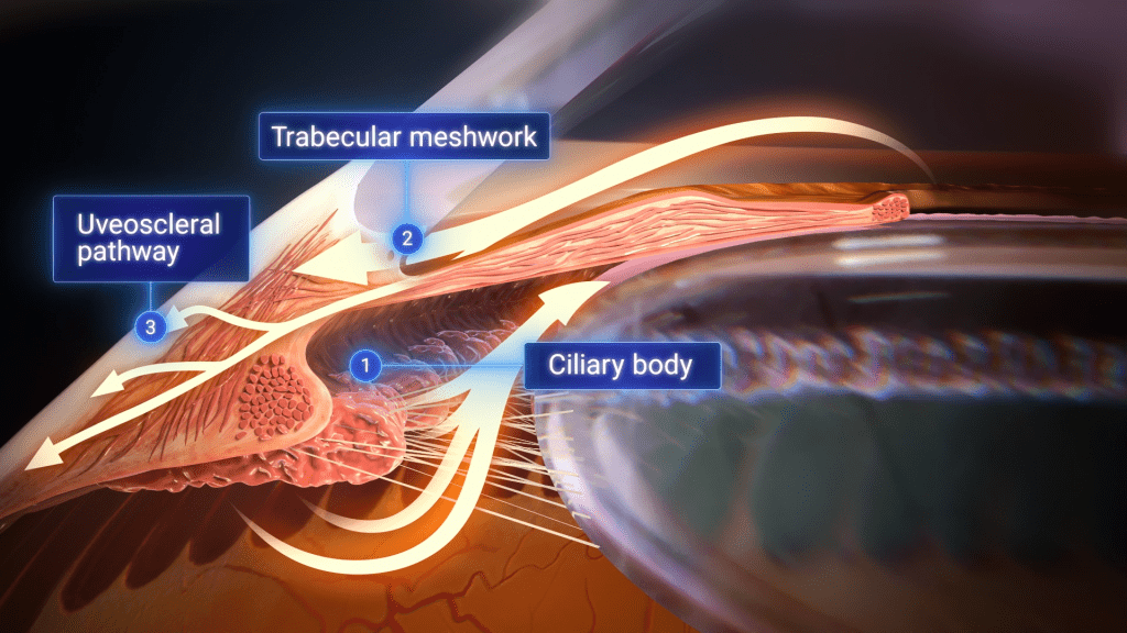
QLS-101 has a markedly different mechanism of action than currently available therapies that target increased eye pressure. Namely QLS-101 is designed to widen vessels distal to the trabecular meshwork of the eye, including the episcleral veins, thereby greatly reducing the resistance in these vessels to lower what is called Episcleral Venous Pressure, which is the largest component of the pressure inside the eye.
With this dynamic and detailed animation, healthcare providers, patients, and investors can better understand how eye pressure is regulated and how Qlaris Bio is attempting to innovate in the field and address an unmet need in targeting this unique component of the pressure inside the eye.