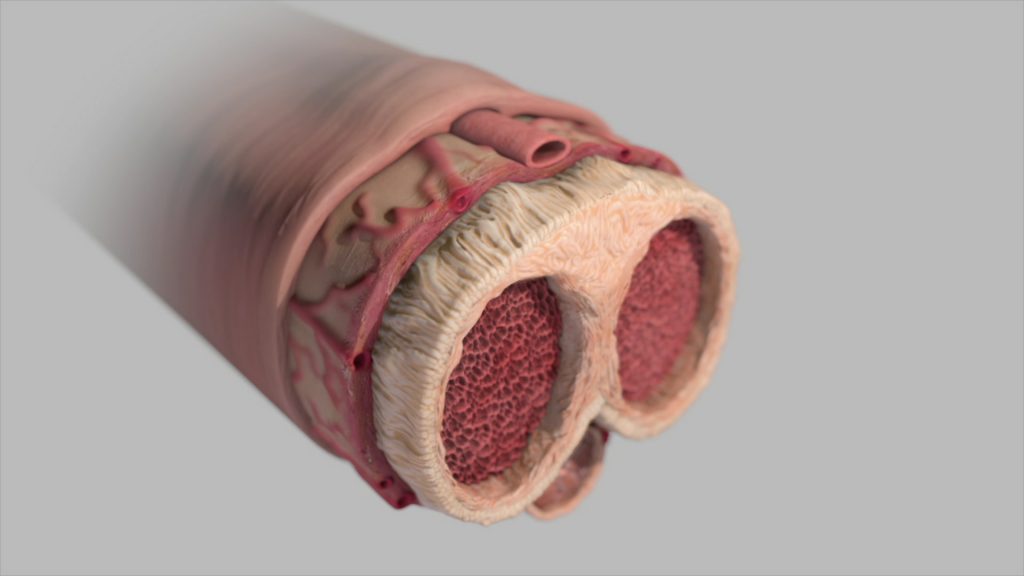
Peyronie’s disease is caused by plaque in the penile tissue. The light beige tissue is the tunica albuginea. It surrounds the corpora cavernosa, and in this condition, it is chronically inflamed.
Related Illustrations
Peyronie’s disease is caused by plaque in the penile tissue. The light beige tissue is the tunica albuginea. It surrounds the corpora cavernosa, and in this condition, it is chronically inflamed.