Skip to content
The Inner Life of the Cell: Protein Packing Animation

Harvard University and XVIVO come together again to add to the growing series of scientific animations for BioVisions – Harvard’s multimedia lab in the department of Molecular and Cellular Biology.

An abstract, colorful 3D illustration depicts a virus with a spherical, textured surface in blue and purple, surrounded by clusters of yellow and orange protein-like structures, capturing the intricate beauty of cell protein packing against a blurred background.

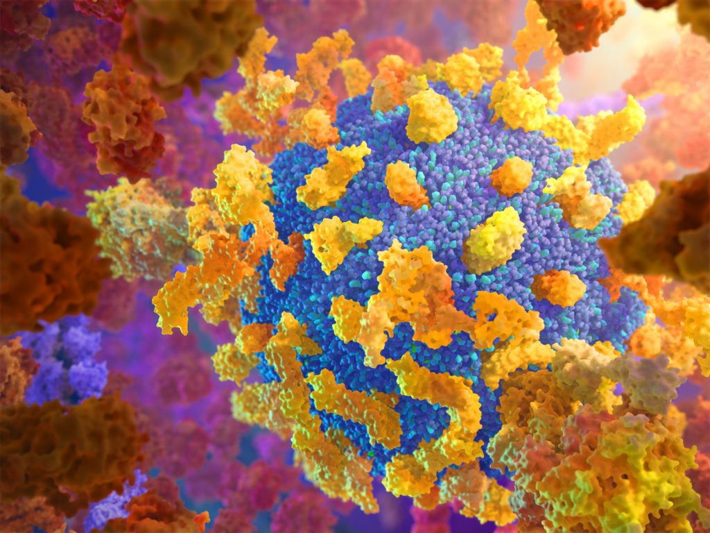
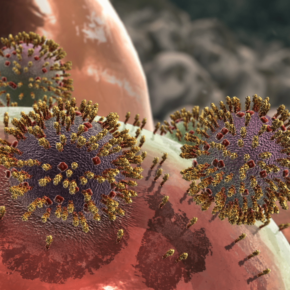
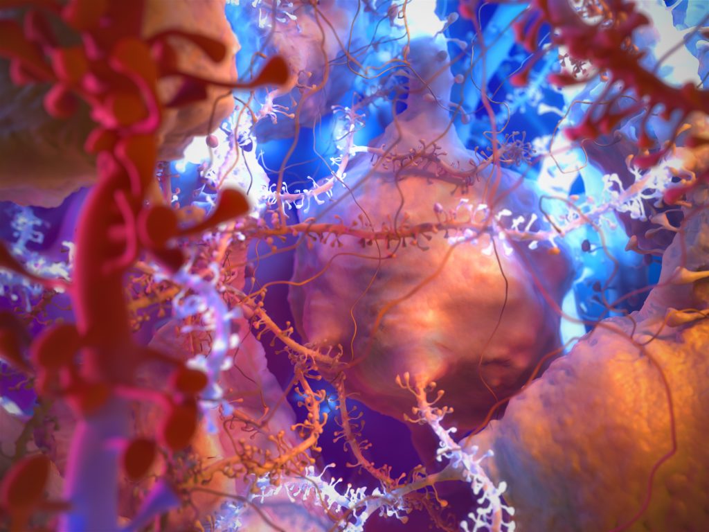
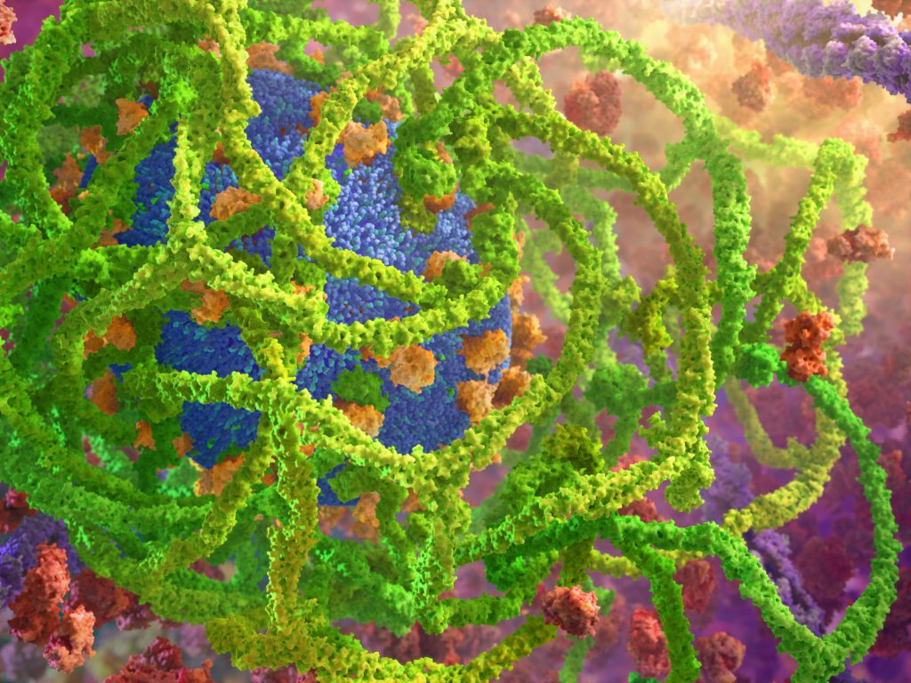
Protein Packing strives to more accurately depict the molecular chaos in each and every cell, with proteins jittering around in what may seem like random motion.

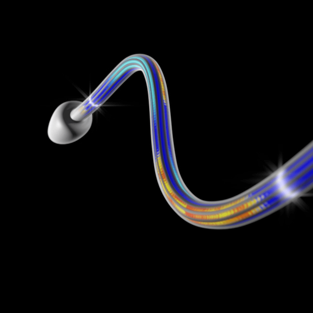
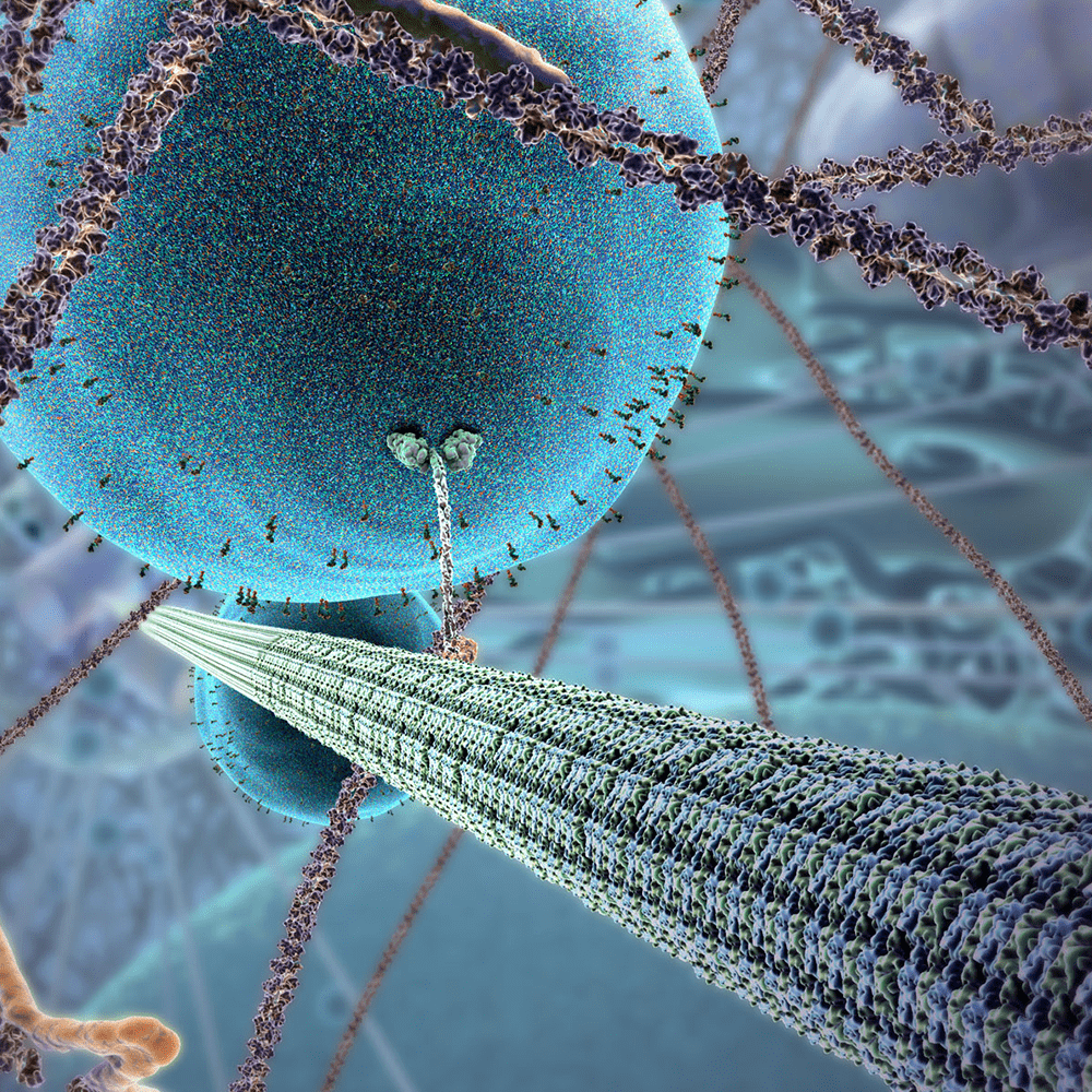
Proteins occupy roughly 40% of the cytoplasm, creating an environment that risks unintentional interaction and aggregation. Via diffusion and motor protein transport, these molecules are directed to sites where they are needed.

A vibrant close-up illustration of nerve cells with pink neurites and synapses against a blue background. The image intricately details protein-packed neural networks and cell bodies in an abstract, colorful style.
An abstract, colorful 3D rendering of a molecular structure showcases the proteins intricate design. Green, twisted strands surround clusters of blue spheres and orange particles, evoking the complex packing within a cell, set against a blurred, multicolored background.

View the first two of this 3D animation series, The Inner Life of the Cell, and Powering the Cell: Mitochondria, on our website.

Related Animations

LIFE CYCLE OF INFLUENZA A – Narrated by: Forest Whitaker

LIFE CYCLE OF INFLUENZA A – Narrated by: Forest Whitaker

View
How Do Cilia and Flagella Move?

How Do Cilia and Flagella Move?

View
THE INNER LIFE OF THE CELL

THE INNER LIFE OF THE CELL

View