The Vaccine Makers Project, a program of the Vaccine Education Center at Children’s Hospital of Philadelphia (VEC@CHOP) and Medical History Pictures (MHP), partnered up again with XVIVO to help develop two new medical animations, this time about how COVID-19 mRNA and viral vector vaccines work.
Here, we’ll show you what went on behind the scenes to make the COVID-19 vaccine animations.
From the start, our Creative Director, KC Knack, knew our animation needed to be accessible to the public audience, with accurate science presented in an approachable format. And all within a 2-minute video.
Vaccine Makers Project provided XVIVO with a draft script that served as our foundation of what to visualize in the animation. With that in hand, our illustrators got to work researching and sketching ideas for the visuals in the storyboard phase. Dozens of hours go into the research, storyboard, and each scene of animation.
Due to the continuous emergence of new published data regarding both SARS-CoV-2 and the COVID-19 vaccine types represented in these animations, there were several characters, processes and environments visualized which were based on a combination of the relevant data available at the time of production, as well as an understanding of both software and hardware limitations.
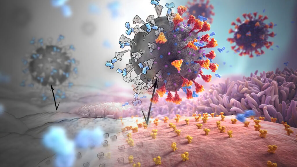
A visual representation challenge we faced, was that of the virus itself. Because SARS-CoV-2 was a novel coronavirus, the precise structure of the virus was yet to be completely understood. One of the viral surface proteins of interest, the spike protein, was often represented in publicly available rendered images of the virus without the Heptad Repeat 2 (HR2) region. During the research process the illustrators found several scientific papers which provided images of the complete modeled extra-virion portion of the spike protein including the HR2 region. We incorporated this information in our representation of the spike protein which resulted in a spike protein with a longer stem.
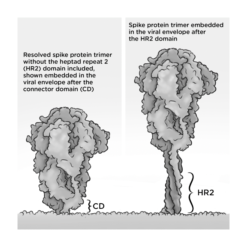
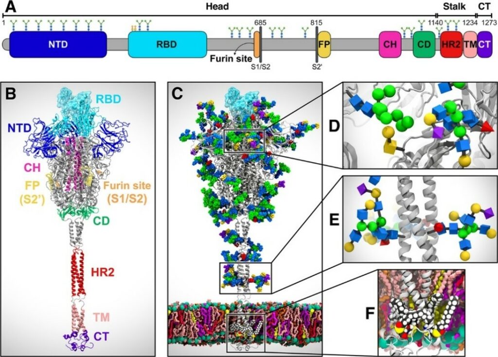
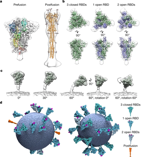
There were many helpful references that we used to inform the modeling of the spike protein in our animation. Below are two of those references.
References used to inform the longer stem structure (including HR2)
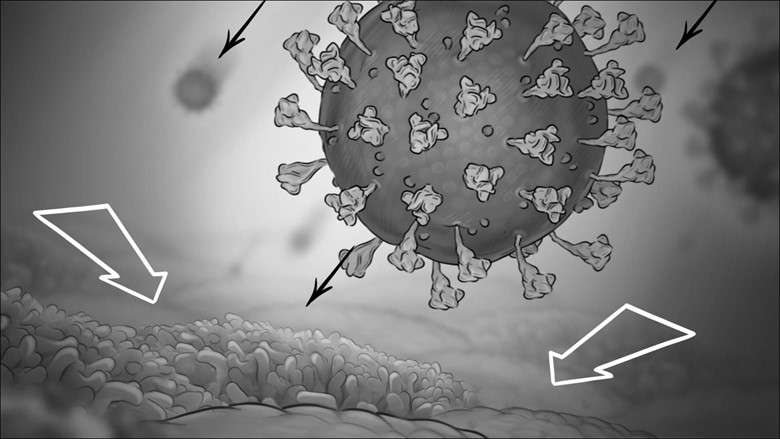
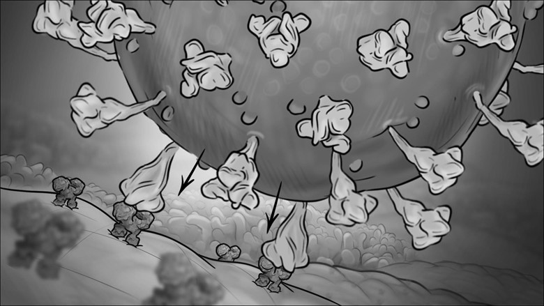
With an approved narration and accurate data on hand, our medical illustrators began storyboard development. These consisted of black-and-white drawings of each scene.
The storyboards detailed everything that eventually takes place in the final program, including voice-over narration, key visual events, and text-on-screen.
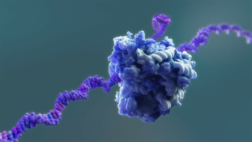
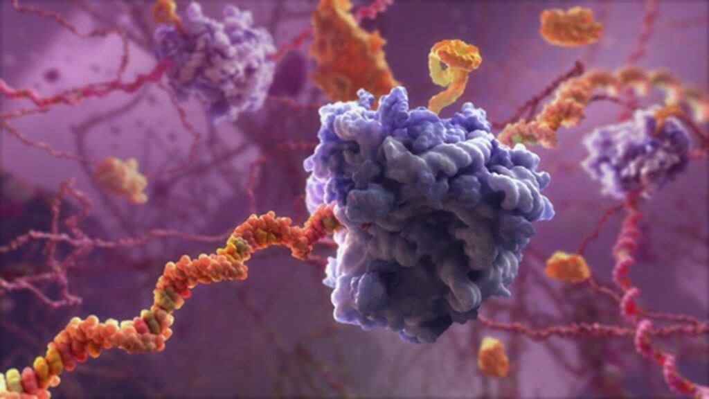
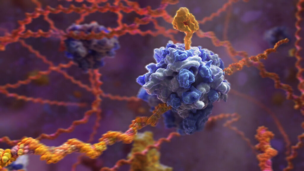
After the narrative and story flow was established in the storyboards, our animators got to work quickly, understanding the timely nature of the project, and started providing visual development excerpts of the proposed animation. Here is a series of visual iterations on the translation of the SARS-CoV-2 spike protein. Our animators developed the translation machinery first, and then through careful lighting and compositing, balanced the colors and values of the background to bring the scene to life.
The animatics and the subsequent animations then went into production, at each stage garnering feedback on the visualizations. This project was animated with, Softimage, Maya, Cinema 4D and Houdini. Our lead animator on this project, Edward Quirk, used his background in movies and extensive animation experience to guide the process.
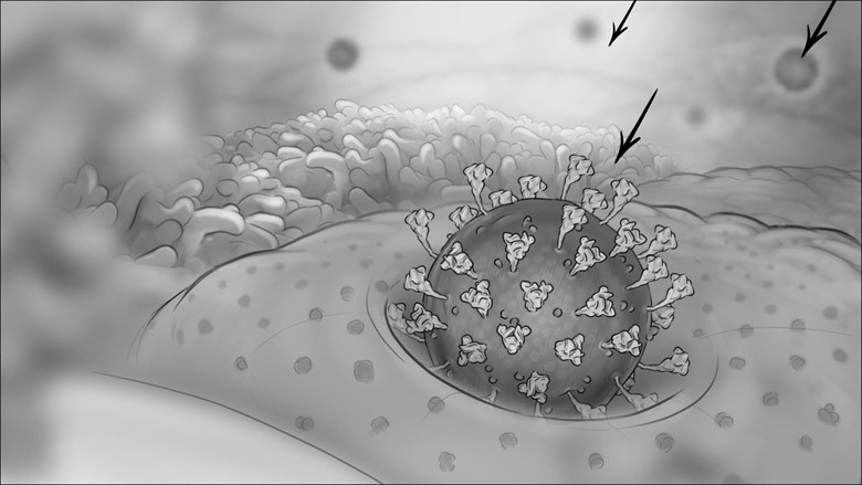
One particular element of the animation that required finesse was the lipid nanoparticle being taken up by the host cells and then the vaccine mRNA being released into the cytoplasm.
To achieve an interaction that looked seamless to the viewer, our animators had to match the geometry of the lipid nanoparticle to that of the membrane, as well as change the layers of lighting, textures, and transparency during the transition. We also had to make educated decisions on some of the scientific aspects of this interaction since certain details regarding the vaccine LNP structure, its cellular uptake and the process of mRNA endosomal escape were not available at the time.
In the end, any animation is a blend of scientific accuracy, visual clarity, and technical requirements to tell a complicated story in an engaging way.
Upon receiving the final animation, the team at Vaccine Makers Project said, “We all agree, this looks awesome!”. Check it out!
XVIVO has a longstanding collaboration with the Vaccine Makers Project, including an earlier 9-part animation series aimed at educating school-age audiences about the immune system and vaccines. The earlier animations provided introductory information on the immune system, as well as viruses and vaccines. In particular, the animations “A virus attacks a cell” and “How do viruses reproduce” have become quite popular during the pandemic.
We were excited when this animation hit social media audiences and became a viral video. Then this tweet appeared from @DrEricDing: “BEST. VIDEO. ALL. YEAR. Please share with friends how the mRNA vaccine works to fight the coronavirus.” We were blown away by how well the video was being received.
We think the success of this piece is due to how a very complex process was distilled into effective visual storytelling, to make the otherwise dense material more easily and intuitively ‘read’ and understood.
We are so inspired by the impact of helping millions of people understand how COVID-19 mRNA and viral vector vaccines work.
Thank you to everyone who has viewed and shared our videos.