![3d microscope animation 1](https://xvivos3bucket.s3.amazonaws.com/wp-content/uploads/2021/03/14151810/NIKON-%E2%80%93-EXPAND-YOUR-VIEW.mp4.00_00_24_17.Still002-1024x576.png)
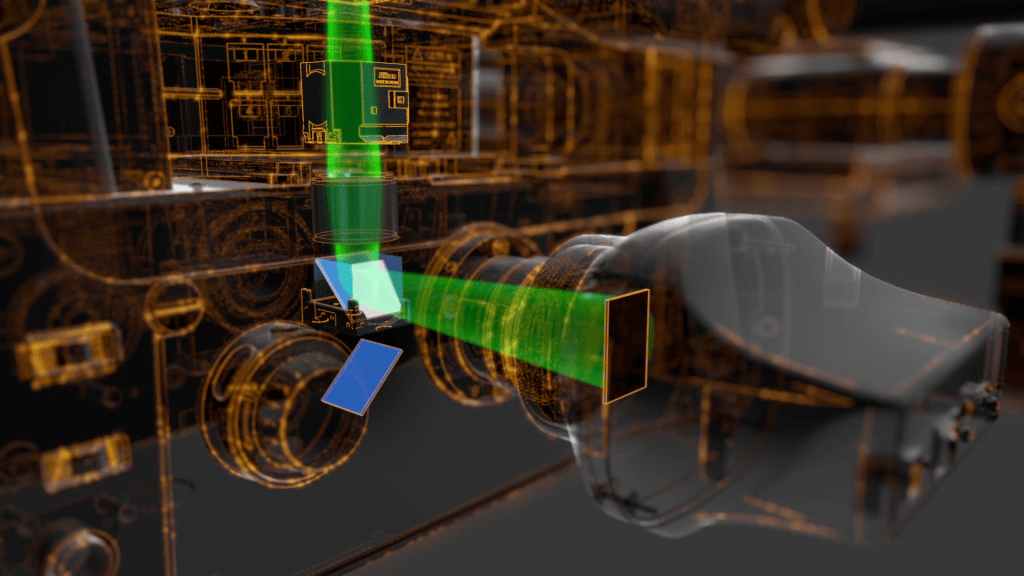
A key part of the engineering that makes the Nikon Eclipse T12 a formidable microscope is the expanded field of view it offers. The second animation in a 4-part series on the new T12 microscope showcases this large field of view, helping the viewer appreciate how the field of double size is attained with a larger lens, and how that translates to a higher throughput of imaging.
![3d microscope animation 2](https://xvivos3bucket.s3.amazonaws.com/wp-content/uploads/2021/03/14151809/NIKON-%E2%80%93-EXPAND-YOUR-VIEW.mp4.00_00_11_10.Still001-1024x576.png)
The semi-transparent effect also shows the inner workings of the microscope in action, which culminates in an actual, beautifully detailed fluorescently-labeled embryo. Learn more about these animations in our blog, NIKON – SEE MORE THAN BEFORE.
Related Animations
![HEART SCIENCES MYOVISTA WAVELET ECG TECHNOLOGY](https://xvivos3bucket.s3.amazonaws.com/wp-content/uploads/2021/02/14151953/HEART-SCIENCES-MYOVISTA-WAVELET-ECG-TECHNOLOGY_Thumbnail_1000x1000.png)
![STRATTICE RECONSTRUCTIVE TISSUE MATRIX](https://xvivos3bucket.s3.amazonaws.com/wp-content/uploads/2021/02/14151953/STRATTICE-RECONSTRUCTIVE-TISSUE-MATRIX_Thumbnail_1000x1000.png)
![GINTUIT™](https://xvivos3bucket.s3.amazonaws.com/wp-content/uploads/2021/02/14151951/GINTUIT_Thumbnail_1000x1000.png)